Granulite Gallery
Petrological features of granulites, charnockites and migmatites
- Outcrop features of granulite-facies migmatites
- Vein-like and patchy charnockite in southern India
- Metamorphic history of the Namaqualand granulites
You may make use of these photographs for bona fide educational purposes. Please acknowledge their source, and let me know what you do with them.
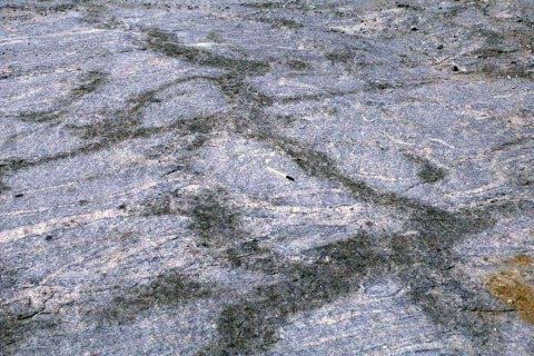
Field photographs of migmatite styles
See also: Waters, D.J. 1988. Partial melting and the formation of granulite facies assemblages in Namaqualand, South Africa. J. Metamorphic Geol., 6, 387- 404.
|
Migmatitic biotite gneiss of semi-pelitic composition. Light-coloured neosome consists of streaks and patches of coarse quartz and feldspar with large garnets. There is no melanosome. The unfoliated neosomes have indistinct boundaries and although they approximately follow the foliation in the rock they are locally discordant. |
|
|
Pink garnet-bearing neosomes are arranged as if they occupy or nucleate around tensional fractures in this metapelitic rock from the central zone of the Damara belt, Namibia. |
|
|
The compositional layering in this metapelite from Namaqualand represents former graded bedding. The neosomes develop around large pre-existing garnets in the more aluminous layers. These rocks are described in Waters & Whales (1984) Contrib. Min. Pet. 88. |
|
|
Neosome in metabasite consisting mostly of very coarse ortho- and clinopyroxene. The neosome has very much less plagioclase than the palaeosome and probably represents the accumulation of solid products of the dehydration melting of hornblende, the melt fraction having escaped. |
|
|
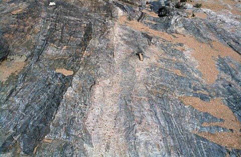
Network of interconnected neosomes in metabasite, indicating how pathways may develop for melt extraction. |
|
|
Pale sheets of granitoid material with sparsely-distributed garnets, approximately parallel to the regional foliation probably represent the extracted melt from nearby metapelites. |
|
|
|
|
|
Melt which crystallises in place will release its water content (originally around 5% by weight at 750°C, 5kbar). If this occurs in contact with the anhydrous solid products of melting, the dehydration-melting reactions may partially reverse themselves. Here biotite + sillimanite + quartz aggregates occupy embayments in a neosome garnet. |
|
|
|
|
Vein-like and patchy charnockite in southern India
|
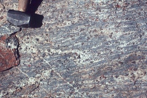
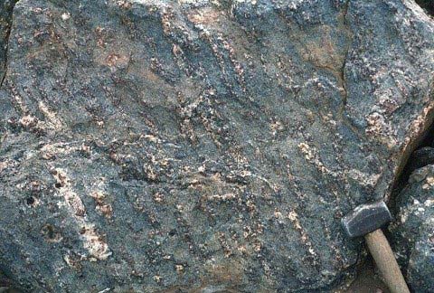
Dark charnockite forms veinlets and patches obliterating the structure of granodioritic Hbl-Bt gneiss in Kabbaldurga quarry, South India. |
|
|
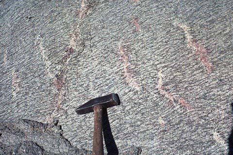
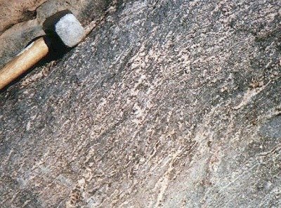
Detail of charnockite vein overprinting foliation in Hbl-Bt gneiss, Ponmudi, South India. |
|
|
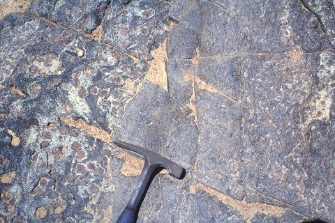
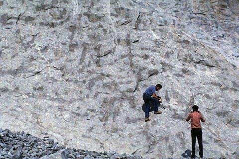
Charnockite patches make a patterned array across the wall of the quarry at Kottavattom, South India. |
|
Metamorphic history of the Namaqualand granulites
|
A view of one of the critical field areas in northern Namaqualand, where petrographic evidence is preserved for the prograde evolution of the granulite-facies rocks. The sequence of mineral assemblages suggests an anticlockwise path on a P-T diagram. |
|
Timing of metamorphism and deformation
Despite the very high grade of the Namaqualand granulites, there is enough petrographic evidence to indicate that the peak granulite assemblages and dehydration-melting features entirely post-date the major regional compressive deformation D2, and are approximately coeval with upright, ductile, but relatively low-strain deformation D3. See also:
- Waters D.J. (1990) Thermal history and tectonic setting of the Namaqualand granulites, Southern Africa: clues to Proterozoic crustal development. In "Granulites and Crustal Evolution", eds. D. Vielzeuf and P. Vidal, NATO ASI Series C-311, Kluwer, Dordrecht, 243-256.
- Waters D.J. (1989) Metamorphic evidence for the heating and cooling path of Namaqualand granulites. In "Evolution of Metamorphic Belts", eds. Daly J.S., Cliff R.A. and Yardley B.W.D., Geol. Soc. London Special Publication 43, 357-363.
|
The present compositional layering in this aluminous gneiss, and the fabric defined by very coarse sillimanite prisms on the left, runs NNW in the photograph. Inside the cordierite porphyroblast at the centre, however, an earlier E-W fabric defined by small sillimanite needles is still visible. Inclusion fabrics elsewhere indicate that this early fabric developed when the rock was a biotite-sillimanite schist. Now its assemblage is hercynite (black) + cordierite + sillimanite + K-feldspar + quartz. |
|
|
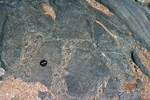
Undeformed garnet + K-feldspar migmatite leucosomes follow the steep-plunging L2 lineation. This does not signify syn-D2 melting: the lineation is defined by a rodding of compositionally distinct domains, in which later melting has preferentially occurred. |
|
|
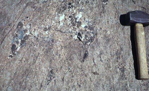
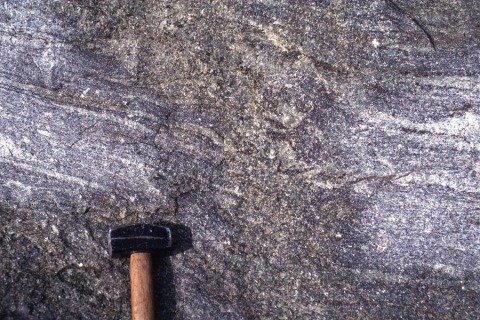
Compositional layering in metapelitic gneiss, parallel to the hammer shaft, is cut by an array of narrow planar migmatite leucosomes, dipping more steeply to the left. The leucosomes define a fabric axial-planar to local F3 folds. |
|
Amphibolite-facies minerals replaced by granulite-facies intergrowths
See also: Waters D.J. (1986) Metamorphic history of sapphirine-bearing and related magnesian gneisses from Namaqualand, South Africa. J. Petrol. 27, 541-565
|
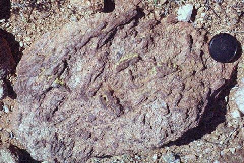
Pencil-shaped porphyroblasts stand out on the surface of this cordierite-phlogopite gneiss. In detail they are prisms with square cross-section, reminiscent of andalusite. |
|
|
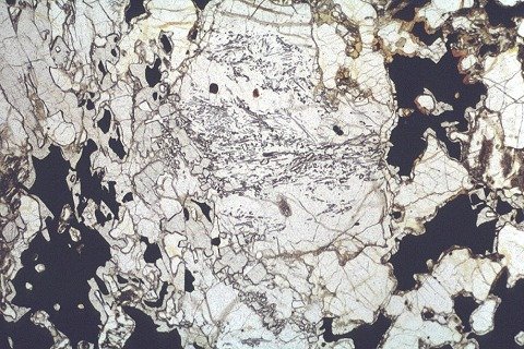
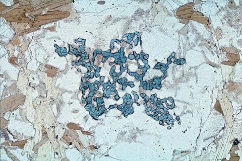
In thin section the porphyroblast shapes are composed of an intergrowth of blue sapphirine with colourless cordierite. |
|
|
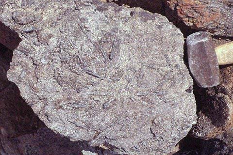
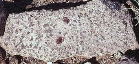
Large, dark, bladed amphibole-like "porphyroblasts" stand out on the surface of this plagioclase-rich gneiss. |
|
|
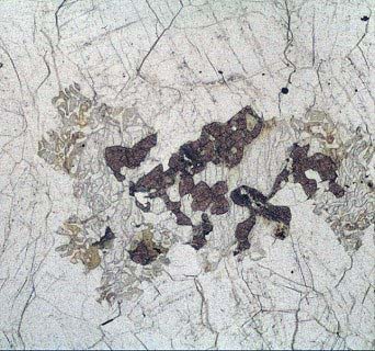
In thin section they are intergrowths of orthopyroxene with cordierite. |
|
|
In this rock the dark patches (up to 2 cm across) are rectangles and lozenges which mimic the crystal habit of staurolite. In thin section (no picture) they are spinel + cordierite nodules. |
|
|
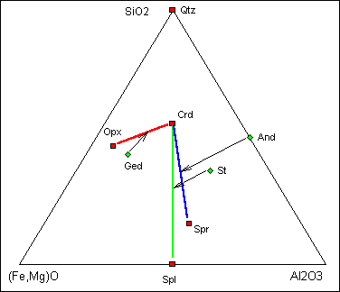
This diagram reveals what happened. The amphibolite-facies precursor minerals (green diamonds) were replaced by pairs of intergrown granulite-facies minerals (red squares). The transformations, shown by the black arrows, are not isochemical, but the proportions of the products are consistent with a replacement process which locally conserves Al and Si. These are expected to be the least mobile components in the absence of aqueous fluid at granulite-facies conditions. |
|
Content last modified March 2000