| Home | Geological History | Stratigraphy | Area map | Rock Index | About |
|
[ About the site ] |
[ About jargon ] |
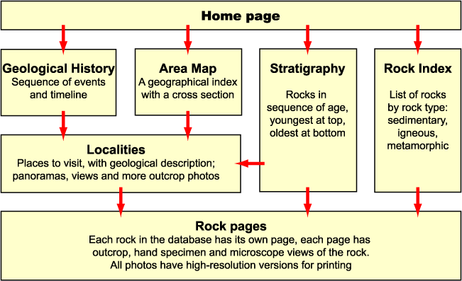
This diagram shows the routes you can take through the explanatory material to the rock images, and there are active links to the main sections and to examples of locality and rock pages.

The high resolution images are, I'm afraid, not available via the web-delivered version of the collection.
The Oxford University Department of Earth Sciences runs a field course for first-year students based at the Assynt Field Centre, Inchnadamph, near Lochinver, Sutherland. This is the students' first real encounter with rocks in their regional context: they learn to describe them, to map out their relationships, and to deduce their origins. We have sampled the rocks in the field, so that we can use hand specimens and thin sections of most of the typical rock types for laboratory studies both in the Assynt field centre and back in Oxford. We have found it useful to have images of the material, both digital and printed, for a number of purposes. Views of the microscopic textures can be taken into the field, for discussion on the outcrop. Field views, along with other physical and virtual material, can be used for pre-course familiarization and post-course consolidation and revision.
The rock types of NW Scotland make quite good general examples, comprising a wide range of sedimentary, igneous and metamorphic rocks. So, when we decided to support Earth Science teaching in the National Curriculum, it made sense to build on materials we were already developing. Moreover, as well as illustrating the processes by which rocks form, they have a fascinating geological and historical context. These rocks tell us about the changing geological environments over almost three thousand million years of Earth history, and they have played a crucial role in the development of geological thinking over the past century and a half of the human era. We hope you will find the images, and the story linking them, useful and informative.
Every scientific discipline has its special language of technical terms. This is quite healthy: the special words have precisely-defined meanings that are understood in the same way by all whose business it is to work in that area. However, to an untrained outsider, the language can look like impenetrable jargon.
We have tried to minimize the number of technical terms in the text, but some are unavoidable. Many can be found in a good general dictionary. As many as possible are defined where they occur, and for others the meaning may in any case be apparent from the context. Later, we hope to add a glossary.
The study of rocks under the microscope yields an abundance of information about their texture and the processes that formed them. Although most rocks are effectively opaque in hand specimen, most rock-forming minerals are transparent when cut thin enough. They are studied in thin section: a small rectangular slab about 3 cm by 2 cm is cut from a hand sample using a diamond saw. It is cemented to a microscope slide, and ground down until it is 0.03 mm thick. After adding a cover slip, it is ready for study in the polarizing microscope.
The microscopes that geologists use have a stage that rotates, and are fitted with polarizing filters, one below the stage and one above. The upper polarizer can be inserted or removed from the light path. The lower one is usually fixed in place.
Light can be considered as a wave motion, vibrating at right angles to its direction of travel. Polarizing filters allow only light vibrating in a particular direction to pass through. Thus, polarizing sunglasses allow only light with up-and-down vibrations into your eyes, cutting out the horizontal vibrations that are associated with reflective glare from water, snow, etc.. Similarly, the lower polarizer allows only left-right vibrating light (plane polarized light) to pass through the thin section.
Viewing a thin section in plane polarized light is much like viewing it in ordinary light, although the appearance or colour of certain minerals may change somewhat as the stage is rotated. Different minerals can be recognised by their colour, relief (how much they appear to stand out), cleavage (regular fractures) and other properties.
The upper polarizer, however, has its allowed vibration direction arranged at right angles to the lower one. This should, when it is inserted, cut out all the light that passes through the lower polarizer. However, most minerals show the property of double refraction. A ray of light passing into such a crystal is split into two rays that are polarized at right angles to each other and travel at different speeds through the crystal (the refractive index is different for the two vibration directions). Light that has been forced into these new directions by passing through a crystal will be able, in part, to pass through the upper polarizer. The amount of light transmitted changes as the microscope stage is turned, the crystal going momentarily dark every 90° when the polarizing directions of the crystal and the microscope polarizers coincide. Moreover, the light transmitted through crossed polars appears coloured, the colour seen depending on the difference between the speeds or refractive indices of the two rays in the crystal. These interference colours not only allow us to distinguish the different grains in an aggregate of minerals, but they are characteristic of the mineral concerned and help us to identify it. Commonly, they reveal other characteristic features, such as twinning in the crystals, that may not be visible in plane polarized light.
|
|
|
Each photomicrograph in the database will indicate in its caption whether it was taken in plane polarized light or between crossed polars. Commonly the same area will be shown in both types of viewing conditions.
| Home | Geological History | Stratigraphy | Area map | Rock Index | About |
D.J. Waters, Department of Earth Sciences, May 2003